
It's time to eliminate your scoliosis pain. Without surgery or injections.
Here at NSC, we’re not just helping people avoid surgeries.
We’re helping people with scoliosis feel whole again.

The Scoliosis Solution® is the #1 exercise-based solution for eliminating scoliosis pain.

“Your program has allowed me to have many days a week with zero pain medication.
This feels like a miracle.”
-Jennifer J.

Our members report an average pain decrease of 31% in three months.

14% of our members report that our non-surgical methods are helping them avoid surgery.
Join Our Free Masterclass
Discover the three secrets that over 25,000 scoliosis patients have used to eliminate their pain from scoliosis without expensive therapies or surgery.

Our Programs
-
![]()
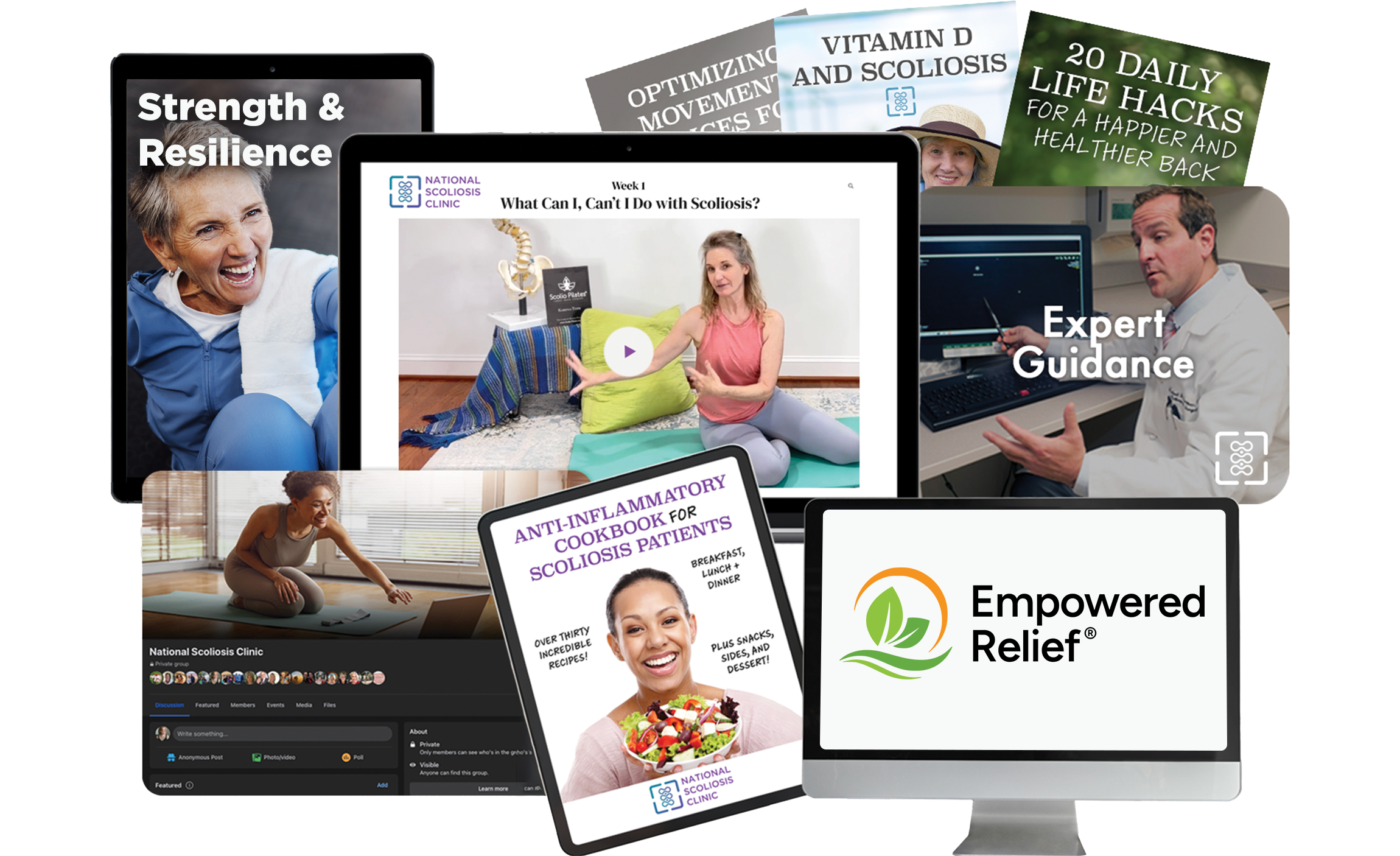
The Scoliosis Solution®
Access our extensive library of scoliosis-safe exercises, eBooks, and pain relief programs. It’s 100% online and on-demand, so you can move through the program at your own pace.
-
![]()
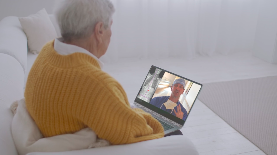
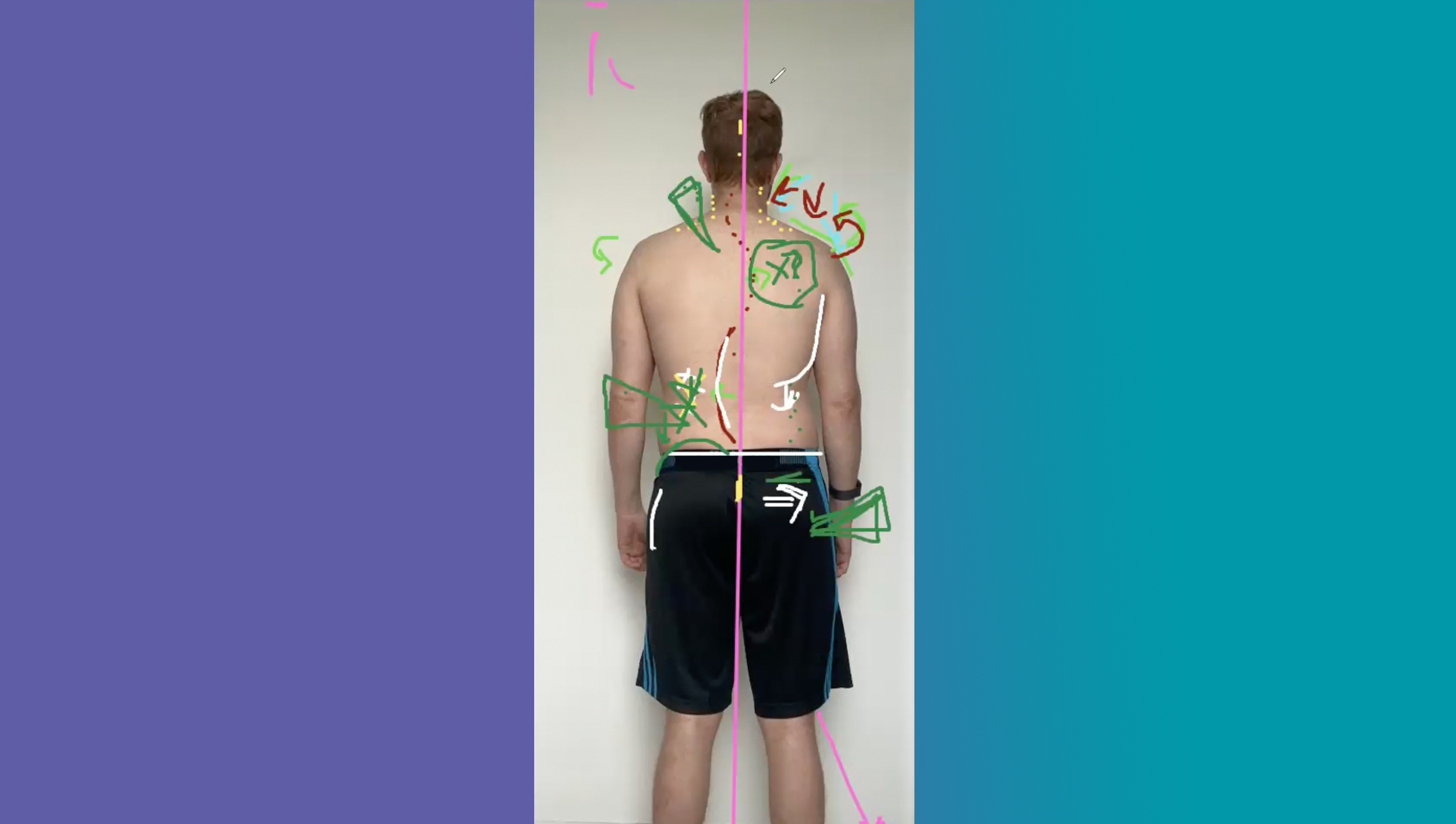
Curved Spine Assessment
Have your spine evaluated by a scoliosis expert and receive a personalized action plan with the movements and exercises that are best for your unique curve.
-
![]()
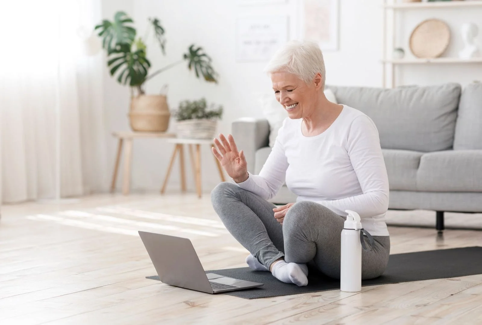
Healing Together Live
Join our online community of people on their journey to a life free from scoliosis pain. Includes office hours, community gatherings, and interactive exercise classes!
Contact
support@nationalscoliosisclinic.com
Reach out to us with any questions, comments, or feedback—your voice matters to us!